Cedar River Clinics is a group of three abortion clinics in Washington that performs abortions all the way past 23 weeks.
On Cedar River Clinics’ abortion page, you will find this direction for women:
If you are unsure about your decision,
-
See the questions and exercises in the online Pregnancy Options workbook.
Pregnancy Options workbook? Sounds informative, right? Well, I headed over to take a look, and here’s what I discovered.
There’s plenty to object to in this workbook, but let’s focus on one disturbing section that is highly misleading and deceptive to women: the fetal development section.
The author provides sketches of preborn babies in their first trimester, next to various pieces of fruit. The author talks about fetal growth in terms of gestational age – the actual age of the baby – not the age of the pregnancy. (For that, add two weeks to the age listed below, since pregnancy is dated two weeks prior to conception, based on the last menstrual period.)

6 weeks gestation (A baby next to a blueberry)

7 weeks gestation (A baby next to a raspberry)

8 weeks gestation (A baby next to a grape)
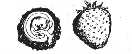
10 weeks gestation (A baby next to a strawberry)
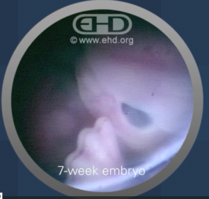
Now let’s look at real images of preborn babies at 6, 7, 8, and 10 weeks gestation – all common ages for abortion. The following images are from The Endowment for Human Development, which has worked with National Geographic to produce a movie about human development in the womb.
6 weeks
EHD has an incredible 40-second video on the baby’s “rapidly growing brain” at this age. You can watch it here. An image from the video demonstrates that a 6-week-old baby also has tiny arms and hands that can move.
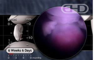
A 4D ultrasound at 6 weeks can be viewed here.
7 weeks

8 weeks
EHD states:
At 8 weeks the brain is highly complex and constitutes almost half of the embryo’s total body weight. Growth continues at an extraordinary rate.
Eight weeks marks the end of the embryonic period. … The embryo now possesses more than 90% of the structures found in adults.
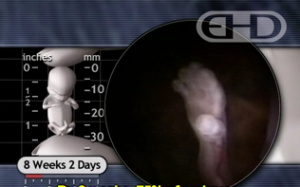
The baby’s waving hand at 8 weeks
10 weeks

“The Sprinter – In a Hurry”
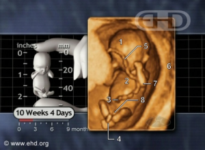
“Hands on Cheeks”

“Crossed Ankles”

“Twins at Play”
Let’s also cover some of the deception – or outright lying – contained in the workbook’s “facts.”
- While the workbook claims the heart changes from a “very small tube” at 7-8 weeks, EHD has footage of a baby’s chambered heart beating at a mere 4 weeks and 4 days. EHD describes it as: “some of the earliest and rarest footage ever obtained showing the separate chambers contract and relax during the cardiac cycle.” See it here. (The heart itself begins beating at 22 days…a fact that the workbook conveniently leaves out.)
- Look back up at the 7-week photos. There are clear eyes, ears, and faces on those babies. Yet, the workbook tells women that from 7-8 weeks “the part of the fetus that will eventually be the face begins to form the shape of eyes and ears.” Eventually be the face? The shape of eyes and ears? Please.
- While the fact is that a preborn baby begins to develop cartilage (a soft skeleton that later hardens to bone) at 5 1/2 weeks, the workbook claims that a soft skeleton doesn’t appear until 9-10 weeks. By that age, bone ossification – the hardening of cartilage to bone – has already been underway for weeks. Ossification starts at 6-7 weeks.
- The workbook claims that, “By the 12th week…[b]lood vessels form in various parts of the fetus and begin to connect to one another.” In reality, by only 8 weeks, the baby’s “scalp has a rich blood supply as does the entire head and neck.” EHD further explains: “The complexity achieved by the embryo in just the first 3 weeks of development is incredible. Considering the importance of distributing nutrients to the emerging brain and spinal cord, as well as the rest of the embryo, the early development of the circulatory system is not surprising. Early red blood cell precursors are present in the yolk sac just three weeks after fertilization! … Also by 3 weeks, early blood vessels form throughout the embryo as the network of the early circulatory system begins to take shape.” So, actually, abortion clinic workbook, blood vessels form before many women even know they’re pregnant!
We could go on. But I’ll stop here, and let you check out more of the true scientific facts from The Endowment for Human Development. After all, who’s more likely to tell the truth – an abortion clinic, or a scientific research organization who exists to “provides a memorable and scientifically accurate framework”?


