Full term 'miracle' baby born after 'unprecedented' ectopic pregnancy
Bridget Sielicki
·
Human Interest·By Amanda Vicinanzo
Spina bifida surgeries in the womb get a little easier with new 3D models
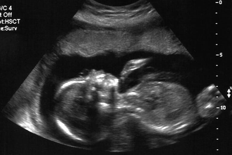
Operating on preborn babies with spina bifida in the womb is not without risk or uncertainty. But that risk has been mitigated with the introduction of lifelike 3D printed models of the tiniest patients to help doctors prepare for in-utero surgeries.
Surgeons at the Orlando Health Winnie Palmer Hospital for Women & Babies are currently using the models to prepare for in-utero surgeries on babies with spina bifida, a birth defect in which a developing baby’s spinal cord fails to develop properly, according to a press release. Spina bifida often leads to walking and mobility issues, and a potential loss of bladder and bowel control. The models were developed by expert 3D printers at Digital Anatomy Simulations for Healthcare, LLC in collaboration with Orlando Health using a combination of MRI, ultrasound imaging, and 3D printing technology.
The realistic models enable surgeons to see details such as skeletal structure, nerve and vascular anatomy, and fluid sacs in the spine and brain caused by spina bifida, giving surgeons the information they need to plan in advance, identify potential obstacles, and even help reduce the duration of the surgery.

“The 3D reconstruction of the fetus can really educate the surgeon on the real-life shape, size and location of the spinal lesion, as well as prepare the surgeon to have the appropriate equipment ready to treat this condition surgically,” Samer Elbabaa, MD, medical director of pediatric neurosurgery at Orlando Health, said in the release. “It’s a level of detail that we are not able to see in traditional imaging, but that is extremely valuable in these cases where we cannot actually see the defect ahead of surgery.”
The models are already giving expecting parents the added layer of confidence they need when approaching surgery.
“At first, we just thought it was a model showing the same kind of condition that our baby was diagnosed with, but then Dr. Elbabaa told us that it was made using the 20-week MRI of our daughter,” Jared Rodriguez said in the release. “We could see the brain and the spine and I looked down at it and thought, ‘I’m holding my daughter right now? That’s pretty awesome.'”
The program may be expanded in the future to include other types of birth defects that can be treated by in-utero surgery.
Article continues below
Dear Reader,
Have you ever wanted to share the miracle of human development with little ones? Live Action is proud to present the "Baby Olivia" board book, which presents the content of Live Action's "Baby Olivia" fetal development video in a fun, new format. It's perfect for helping little minds understand the complex and beautiful process of human development in the womb.
Receive our brand new Baby Olivia board book when you give a one-time gift of $30 or more (or begin a new monthly gift of $15 or more).
READ: Cincinnati hospital reaches groundbreaking milestone in prenatal spina bifida surgeries
While there is no cure for spina bifida, in-utero surgery can yield numerous benefits. According to a study that appeared in JAMA Pediatrics, National Institute of Child Health and Human Development researchers discovered that children who underwent in-utero surgery to repair spina bifida are more likely than those who received postnatal repair to walk independently and perform self-care skills, like using a fork and brushing their teeth.
Live Action News previously reported on several stories of babies who underwent fetal surgery, including Elouise Simpson, one of the first people in the United Kingdom to ever undergo surgery for spina bifida in the womb. Just like other babies, at one-year-old, Elouise took her first steps — a milestone her parents once thought would be impossible for their daughter.
Despite stories like these, many preborn children with spina bifida are aborted. As Live Action News previously noted, a mother of a baby named Lilian, who has spina bifida, claimed she was “offered an abortion at every visit up until 24 weeks.”
These stories serve as a poignant reminder that prenatal surgery and other medical advancements — not abortion — are helping numerous preborn babies around the world with birth defects and disabilities.
“Like” Live Action News on Facebook for more pro-life news and commentary!
Live Action News is pro-life news and commentary from a pro-life perspective.
Contact editor@liveaction.org for questions, corrections, or if you are seeking permission to reprint any Live Action News content.
Guest Articles: To submit a guest article to Live Action News, email editor@liveaction.org with an attached Word document of 800-1000 words. Please also attach any photos relevant to your submission if applicable. If your submission is accepted for publication, you will be notified within three weeks. Guest articles are not compensated (see our Open License Agreement). Thank you for your interest in Live Action News!

Bridget Sielicki
·
Human Interest
Bridget Sielicki
·
Human Interest
Nancy Flanders
·
Human Interest
Nancy Flanders
·
Human Interest
Nancy Flanders
·
Pop Culture
Cassy Cooke
·
Human Rights
Amanda Vicinanzo
·
Human Interest
Amanda Vicinanzo
·
International
Amanda Vicinanzo
·
Issues
Amanda Vicinanzo
·
Newsbreak
Amanda Vicinanzo
·